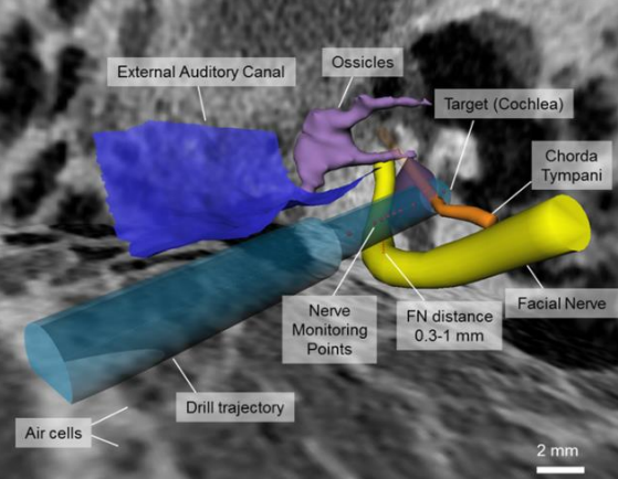
Reported studies pertaining to needle guidance suggest that tissue impedance available from neuromonitoring systems can be used to discriminate nerve tissue proximity. In this pilot study, the existence of a relationship between intraoperative electrical impedance and tissue density, estimated from computer tomography (CT) images, is evaluated in the mastoid bone of in vivo sheep. In five subjects, nine trajectories were drilled using an image-guided surgical robot. Per trajectory, five measurement points near the facial nerve were accessed and electrical impedance was measured (≤1 KHz) using a multipolar electrode probe. Micro-CT was used postoperatively to measure the distances from the drilled trajectories to the facial nerve. Tissue density was determined from coregistered preoperative CT images and, following sensitivity field modeling of the measuring tip, tissue resistivity was calculated. The relationship between impedance and density was determined for 29 trajectories passing or intersecting the facial nerve. A monotonic decrease in impedance magnitude was observed in all trajectories with a drill axis intersecting the facial nerve. Mean tissue densities intersecting with the facial nerve (971-1161 HU) were different (p <;0.01) from those along safe trajectories passing the nerve (1194-1449 HU). However, mean resistivity values of trajectories intersecting the facial nerve (14-24 Ωm) were similar to those of safe passing trajectories (17-23 Ωm). The determined relationship between tissue density and electrical impedance during neuromonitoring of the facial nerve suggests that impedance spectroscopy may be used to increase the accuracy of tissue discrimination, and ultimately improve nerve safety distance assessment in the future.
Electrical Impedance to Assess Facial Nerve Proximity During Robotic Cochlear Implantation


