


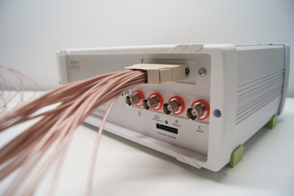

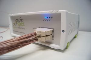


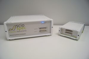


reference systems
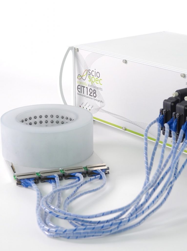
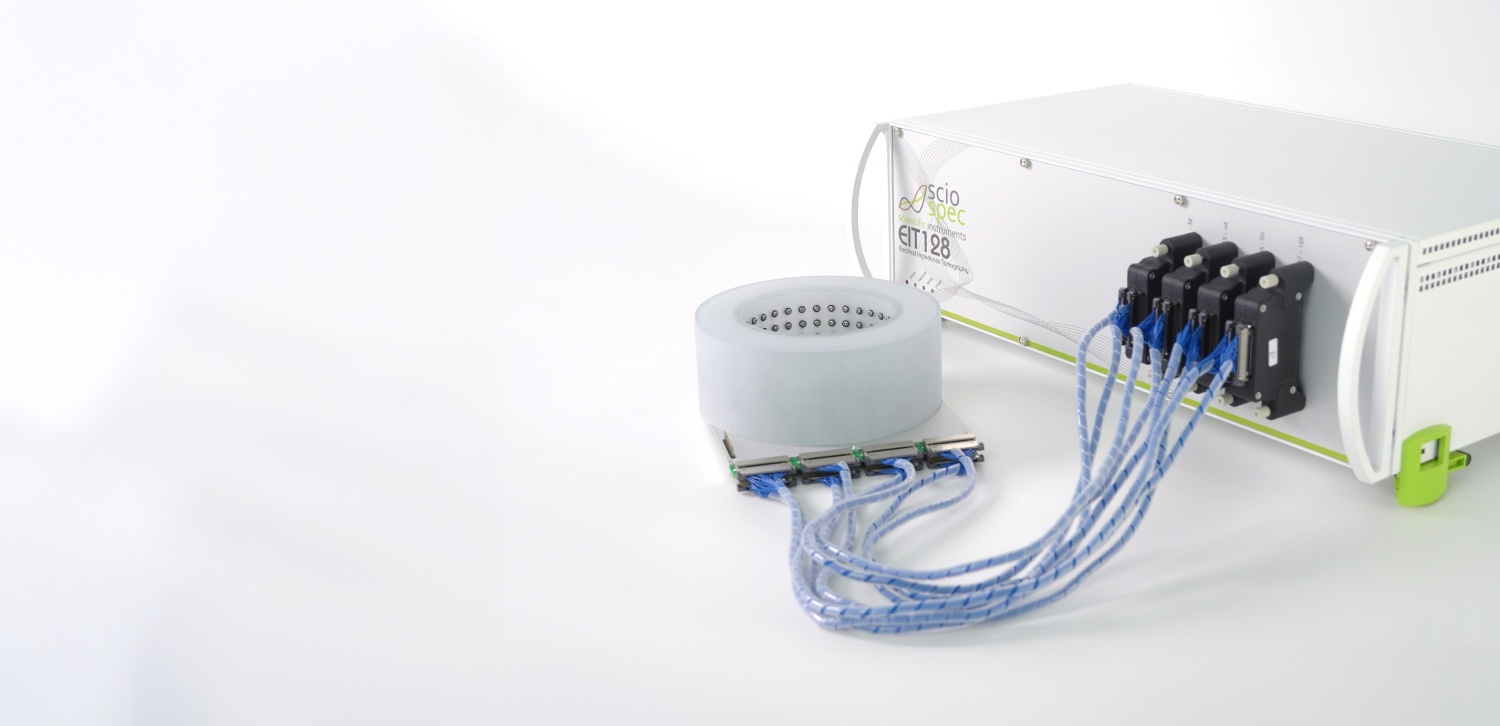
Wassergehaltsbestimmung von tablettierten Pharmazeutika
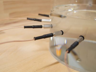
Der Wassergehalt von Pharmazeutika ist ein entscheidender Faktor für viele Aspekte in der pharmazeutischen Industrie. Er beeinflusst unter anderem Funktion, Stabilität, Qualität, Haltbarkeit und Lagerfähigkeit