Skip to content
Home
Products
Impedance Analyzers
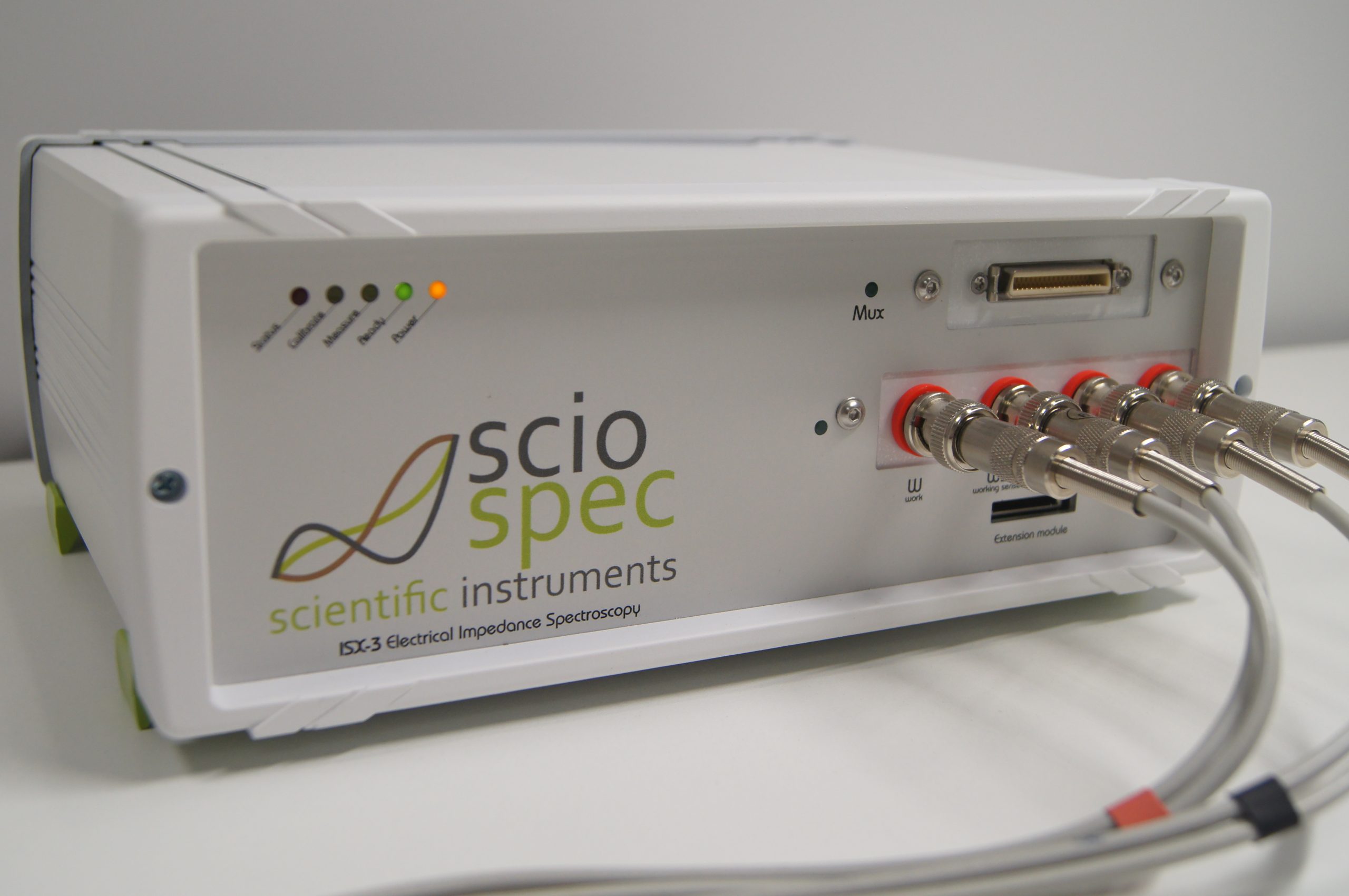
ISX-3
Medical Research ISX-3
ISX-3 EIT
ISX-3mini
ISX-5
CSX-64
LCR Meter
Potentiostat/Galvanotstat
Overview
Electrical Impedance Tomography
EIT16
EIT32/64/128+
LungEIT Kit
ISX-3 EIT
CSX-64
Sensor & Chipadapters
MEArack
ECISadapter
Multiplexer
OEM
References
Request a Quote
About us
English
Support
Search
Contact us
Menu
Home
Products
Impedance Analyzers
ISX-3
Medical Research ISX-3
ISX-3 EIT
ISX-3mini
ISX-5
CSX-64
LCR Meter
Potentiostat/Galvanotstat
Overview
Electrical Impedance Tomography
EIT16
EIT32/64/128+
LungEIT Kit
ISX-3 EIT
CSX-64
Sensor & Chipadapters
MEArack
ECISadapter
Multiplexer
OEM
References
Request a Quote
About us
English
Support
Search
Contact us
FLEXIBLE. PRECISE. AFFORDABLE
Sciospec’s Portfolio
Order By
Publication date
Title
Filter by
Type of reference
application note
Conference
publication
reference systems
research project
whitepaper
Tags
3D cell culture
3D Label-Free Monitoring
96-well-multielectrode array
application-specific
Automated Test
bacteria
Battery
battery-powered
Biofilm
Biofilm monitoring
bioimpedance
Biopsy
biosensor
C#
cancer research
CardioEpiX
cardiology
Cardiomyocyte
Cardiotoxicity
cell migration
cell viability
cell-dependent impedimetric parameters
cell–electrode interface
cement
cementitious materials
coating process monitoring
complex stimulation
conductivity image reconstruction
custom
Dielectric cell model
Dielectric relaxation
ECT
EFP
EIDORS
EIS
EIT
EIT-on-Chip
electrical impedance spectroscopy
Electrochemistry
electrode impedance
Electrophoresis
electroporation
EpiX
EPR
Equivalent circuit modeling
espectroscopia de bioimpedancia eléctrica
ex vivo
FER
fidelity-embedded regularization
flexible sensor
Fluidic dielectrophoresis
Freeze-drying
frequency-difference imaging
functionalised substrates
Galvanostat
geoelectric
geophysical mapping
Glioma
glucose
human heart
Ice
image reconstruction
imaging
Impedance
impedance cardiography
Impedance spectroscopy
Impedance Tomography
impedimetric sensor
in vitr
in vivo
in-vitro
inflammation
Interdigital electrode array
Interdigital electrode arrays
intracerebral electrodes
intraoperative
ISX-3
ISX-3 mini
ISX-5
Lab-on-Chip
Label-free activation monitoring
Leukocytes
lung monitoring
lung ventilation
Lyophilization
massive-multichannel
mathematical modelling
MATLAB
Maxwell-Wagner
MEA
meat
meat processing
Microcavity array
microelectrode array
Microfluidics
mobile health
mPGstat
multi-array impedance sensor
multi-parametric sensor
multichannel
multiwell
needle guidance
NETs
Neuromodulation
neuronal differentiation
neuronal interface
Neuropeptide Y receptor
neutrophil extracellular traps
OEM
operating room
PEF
phantom experiment
pharma
pharmaceutica
Polarization
portable
Potentiostat
pre-clinical testing
pressure sensor
process monitoring
Process-analytical-technology
Pulsed electric fields
QCM
quartz crystal microbalance
real-time bioimpedance
real-time image reconstruction
real-time monitoring
real-time monitoring system
research
Sciospec EIT16
Sciospec EIT8
surgery
Tank Experiment
Through vial impedance spectroscopy
Time-difference electrical impedance tomography
time-difference imaging
Time-resolved quantification of ion channel activity
tissue-discrimination
TRPV1 channel activation monitoring
TVIS
viscosity
water content
wound healing
wound healing detection
wound infection
Wound sensor
See more
Load more
Search
Search
Get in touch with us
Name
Company or institution
Email
Subject
Message
I have read and agree to the
Privacy Policy
Contact us